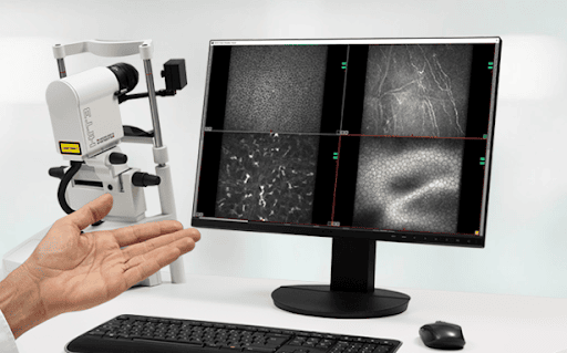
In Vivo Corneal Microscopy (IVCM) is a non-invasive imaging technique based on confocal imaging using near infrared light. Images acquired at the cornea generate a confocal “stack” of cellular level images that comprise different corneal layers. Because the cornea is the most densely innervated area of the human body, there is significant clinical utility from this unique insight into the health of the central nervous system.
Of the tissue layers that are imaged, the sub-basal plexus is of most interest given the density of nerve fibers that are clearly resolved. Their lengths, density and shape have been shown as effective biomarkers in a wide variety of pathologies, including dry eye, acute ischemic stroke, diabetic peripheral neuropathy, mild cognitive impairment and dementia.
As with any imaging-based biomarker, however, utility is based on the efficacy of automated analysis methods. At our heart, Voxeleron is a computer vision shop where we have developed image analysis and machine learning-based methods for a variety of medical imaging modalities. And this is often done in collaboration with our clinical partners. In this instance, we developed deep learning-based methods for IVCM in conjunction with Johns Hopkins School of Medicine. The back-story here relates actual to a collaboration with their neurology group and optical coherence tomography imaging, but during a visit a meeting with Professor Joe Mankowski resulted in us working on his pre-clinical studies of simian immunodeficiency virus (SIV)-infected pigtailed macaques.
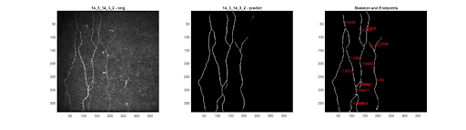
The analysis methods developed automatically measure the lengths of the corneal nerve fibers, their end and branch points, their densities and tortuosity. Since developing these for the pre-clinical models, we have collaborated with other clinical partners, in particular with the University of Auckland, to validate and apply these in the clinical setting in studies including diabetes, colorectal cancer and multiple sclerosis.
Given the uniqueness and complementary information offered by this low-cost modality, there is increasing interest in its use for both ocular and systemic diseases in clinical trials. These biomarkers also contribute to the emerging field of oculomics and are, like our other measurements, extensively validated end points that are available through our iNebula platform.
References
https://www.sciencedirect.com/science/article/pii/S1542012424000752
https://www.mdpi.com/2077-0383/11/16/4770
https://www.nature.com/articles/s41598-021-01226-1
https://journals.lww.com/corneajrnl/Abstract/2021/05000/Combining_In_Vivo_Corneal_Confocal_Microscopy_With.15.aspx
https://eandv.biomedcentral.com/articles/10.1186/s40662-020-00192-5
https://doi.org/10.1101/2020.04.19.048926
https://www.biorxiv.org/content/10.1101/758433v1
http://ajp.amjpathol.org/article/S0002-9440(14)00162-X/fulltext