For a list of ophthalmic publications created by the Voxeleron team, as well as a selection of publications using our software – Orion and Insight – please go to our ophthalmic links page.
User Guides
OrionTM User Guide
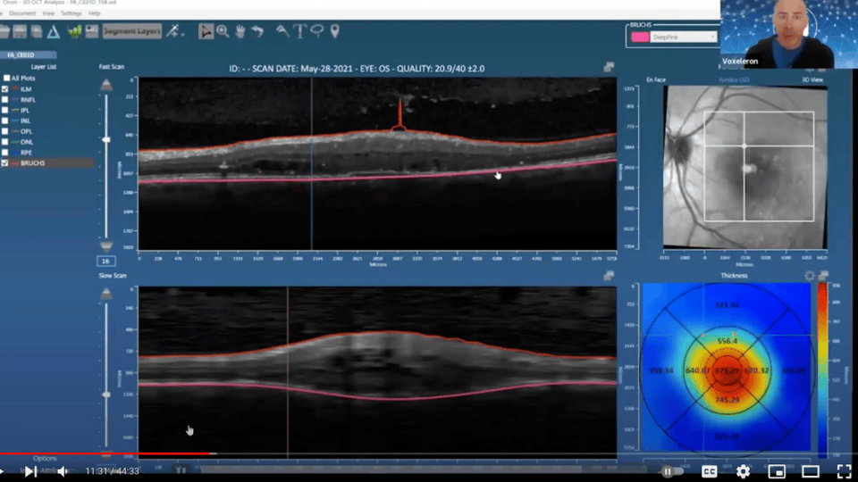
User Guide documenting how to install the Orion software, acquire a license key and then use the software. Covers basic operations, batch processing, editing, 3d viewing, angiography support, adding calipers and annotations, and how to export the results in various formats. It has recently been updated to include the new Change Analysis software.
iNebulaTM Quick Start Guide
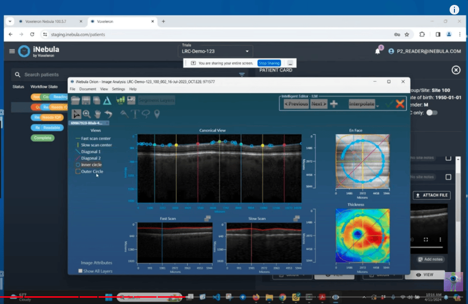
Introduction to using iNebula, Voxeleron’s cloud-based ophthalmic image and data viewing and management system.
iNebulaTM Use Case
Use case to learn how iNebula can ensure effective trial recruitment, management and retrospective analysis.
OrionTM Data Sheet
Data sheet documenting what scan patterns Orion supports, formats and also the install requirements.
OrionTM Notes on Formats
The following gives an overview of how to export Heidelberg Spectralis Data to XML format such that it is readable by Orion.
OrionTM Notes on DICOM Export
And the following gives an overview of how to export Dicom Data from Zeiss’s Forum software for import into Orion.
Pisces 3D – Animal OCT
User guide for our animal OCT analysis software.
Various Reports / Presentations
AAO 2024 Presentation
The following summarizes the results of a multi-center study assessing the performance of our deep learning-based geographic atrophy (GA) segmentation. It assess performance in real-world clinical data from both the Spectralis and Cirrus devices. The subject eyes included were confirmed GA, but also had other comorbidities.
ARVO 2022 Download
ARVO was, as ever, an excellent conference. Relative to our work and clinical trials in particular, we summarized some interesting points raised during special interest group (SIG) meetings where the forums were often quite open due to the ability to more freely interact. These we downloaded in the following.
Blaustein Pain Grand Rounds – 2021
The Blaustein Pain Grand Rounds is a series of lectures delivered as part of The Blaustein Pain Research and Education Endowment. In conjunction with Professor Joseph Mankowski, we presented our collaborative work on nerve fiber segmentation in corneal confocal microscopy images.
Outcome of our SBIR NIH award
The following is the two page summary report that summarizes the results of the specific aims of our SBIR award, grant 1R43TR001890-01, titled: “Retinal image analysis software for neurodegenerative disease research”. Both of the milestones were met. The first was a validation of the segmentation algorithm over 100 diseased eyes from four different OCT manufacturers’ devices. The second related to a validation of the registration algorithms now used in Orion for support of longitudinal analysis.
Algorithms for Clinical Optical Coherence Tomography
In ophthalmology OCT is used to generate in vivo, cross-sectional imagery of ocular tissues. Initially based on time-domain interferometry, the scanners were limited in the amount of data that could be captured within a reasonable time-frame. In the advent of faster, Fourier-domain instruments, OCT scanners are routinely used to acquire truly volumetric image data. As more and more data is acquired, there is an increasing need for automated image analysis tools and reliable quantification. This white paper documents the fundamental clinical OCT algorithms that address these needs.
Regulatory Aspects of Releasing OCT Normative Databases and Algorithms
OCT devices have become the standard of care in the management and detection of retinal diseases. Although OCT devices are not billed as diagnostic devices by their manufacturers, the devices and their algorithms must adhere to strict regulatory procedure. This white paper gives an overview of what is entailed, with particular emphasis on algorithm release and normative databases.
Image Processing for Clinical Optical Coherence Tomography
Johns Hopkins, March 27th, 2012
We have a couple of collaborations at Johns Hopkins, so it was an honor to be invited to talk about one focus of ours, image processing for clinical OCT. Here we were able to meet with neuro-ophthalmologists who are pioneering the clinical use of inner retinal segmentation analyses in OCT.
IEEE EMBS, March 20th, 2013
This talk was an IEEE meeting hosted at Stanford Medical School. The presentation aimed primarily to communicate some of the very exciting recent work in neuro-ophthalmology where retinal segmentation algorithms have been used to support clinical research. Ahead of that, it introduced OCT and the significant role played by image processing algorithms.
Healthcare, Informatics, Imaging and Systems Biology Conference
IBM Almaden, July 29th, 2011
At this conference, we gave a tutorial entitled, “Image Processing for Optical Coherence Tomography”. The following are the slides from that talk.


