Blaustein Pain Grand Rounds Conference October 22, 2021
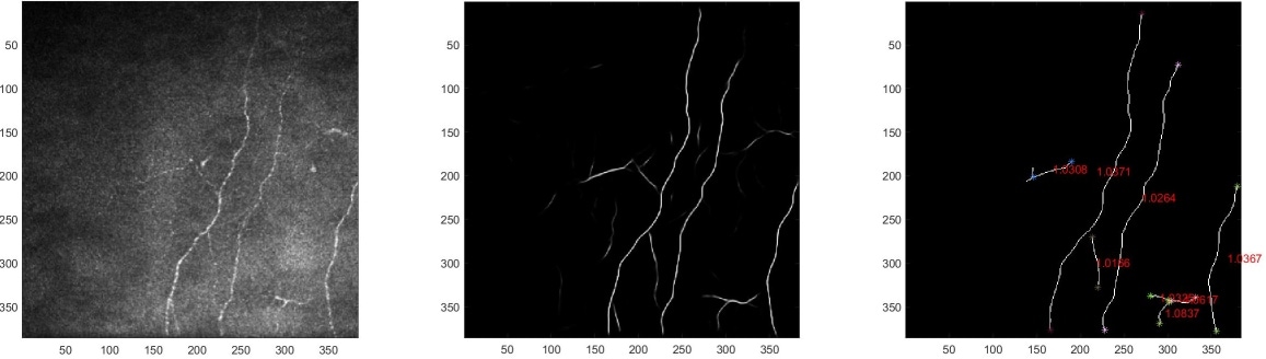
Deep Learning-Based Analysis of Macaque Corneal Sub-Basal Nerve Fibers in In Vivo Confocal Microscopy Images
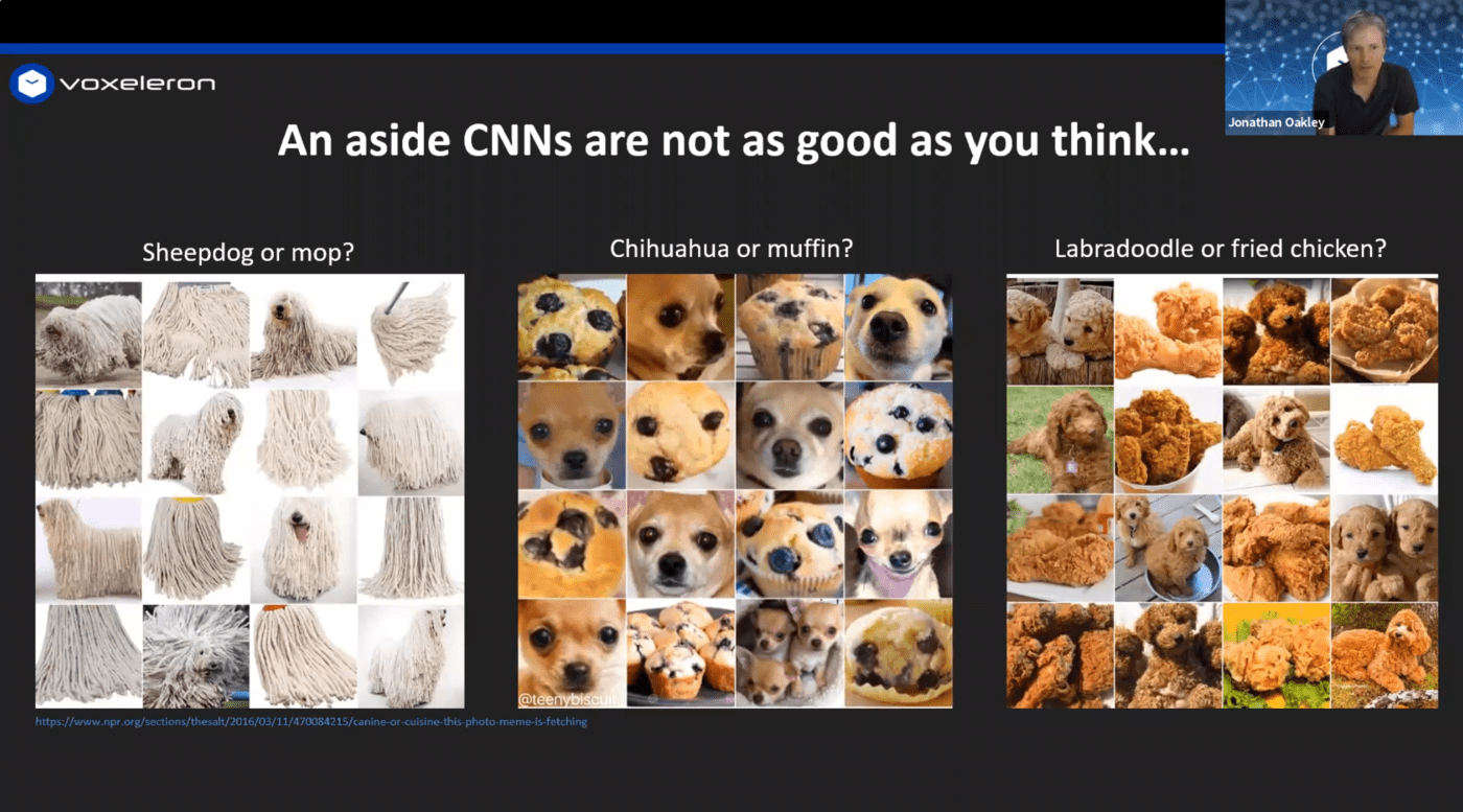
This was a presentation given as part of the Blaustein Pain Grand Rounds Conferences held at Johns Hopkins University School of Medicine. It gives an overview of the collaboration between Prof. Mankowski’s lab and Voxeleron in the automated analysis of macaque corneal sub-basal nerve fibers using confocal microscopy and how the work is being transferred to clinical applications. We also offer some intuition into how convolutional neural networks (CNNs) are actually working as these underpin our approach to automated analysis of the image data.
Passcode: xPHcs!L1