Fluid Quantification in OCT – A Validation using a Home-Based OCT Device
Voxeleron’s AI Retinal Fluid Quantification Highlighted in Retina
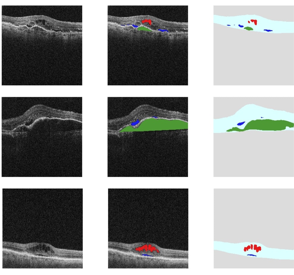
Voxeleron is pleased to announce the publication in Retina of our most recent validation paper on AI-based fluid quantification. In this case it is using the RetinaCheck low-cost optical coherence tomography (OCT) device (OCTHealth LLC, Sacramento, California). This prospective study was based on 70 patients with neovascular age-related macular degeneration (nAMD).
Some Background
In an ideal world, patients with AMD would visit their ophthalmologist at a frequency sufficient to catch early signs of progression to advanced nAMD. Or, if they already have nAMD, frequent visits would be required to ensure proactive management of this disease. But, in both scenarios, only real-time monitoring of patients can truly support optimal interventions. Even before the pandemic, it was a problem following patients at risk for sudden conversion to nAMD closely enough to prevent irreversible vision loss. To make matters worse, the elderly patients most at risk for this disease are often the least mobile, compounding the problem of bringing them into clinics at any kind of regular cadence.
Home monitoring with a low-cost OCT device is one potential solution that would see physicians keeping tabs on their patients remotely ensuring that they are treated in a timely manner. That said, ophthalmologists around the world are already spread very thin and giving them daily images to interpret from each of their remote patients is not a feasible solution. This is why we think our work here is so important. Lower-cost devices, by definition, have cheaper components and must necessarily sacrifice some image quality. We took our existing AI fluid analysis and successfully validated it on such images. Not only is this more challenging than using higher quality images from a clinical device, the size of the study is significantly larger than existing studies based on such clinical data.
The Reported Analysis
The analysis compared our fluid quantification algorithm with hand-segmented ground truth for 136 eyes from 70 patients imaged with a prototype, low-cost home OCT device. For fluid detection our AUC values were 0.951, 0.974, and 0.976 for intra-retinal fluid (IRF), sub-retinal fluid (SRF), and sub-retinal pigment epithelium (RPE) fluid, respectively. For fluid quantification, our R2 model fits for IRF, SRF, and sub-RPE fluid were 0.81, 0.93, and 0.97, respectively. More work is planned, but this is an important foundation for future studies of this prototype home OCT device and highlights its potential to follow high-risk patients outside the clinic. In doing so, it can lessen the disease-monitoring burden for the patients, their caregivers, and the clinic, and also improve visual outcomes for patients with nAMD.