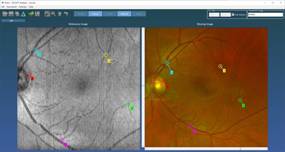
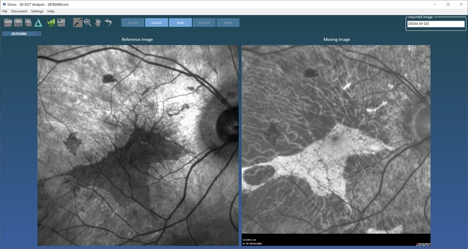
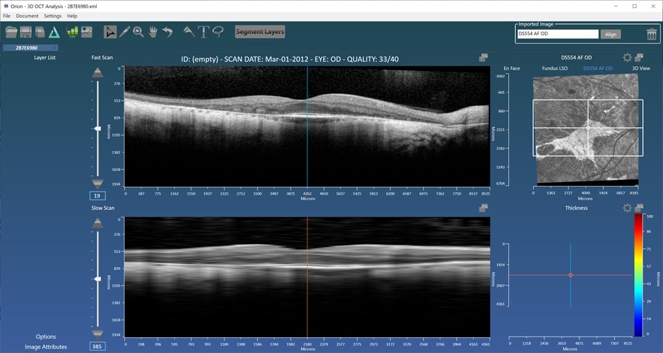
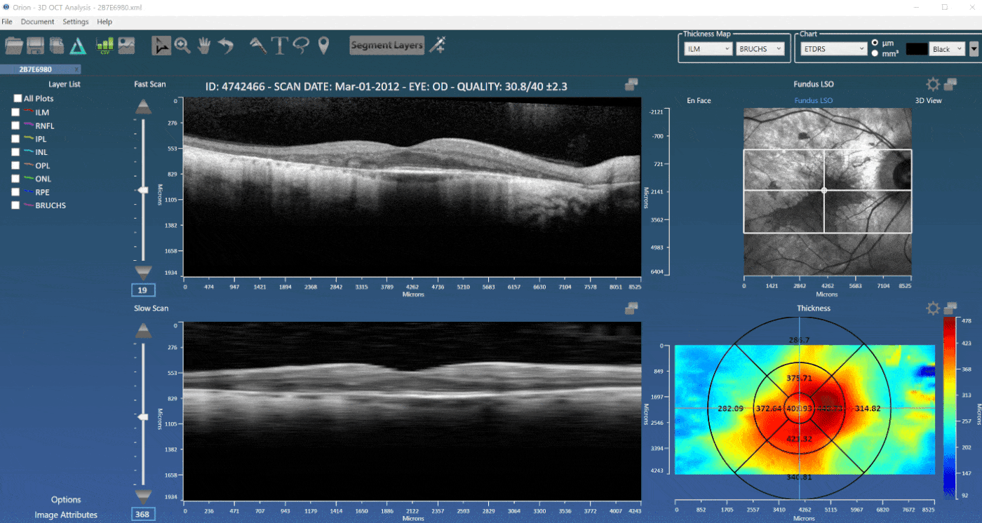
Fundus Image Import
Included in the current release of Orion is the ability to import arbitrary fundus images, be they color fundus images, auto-fluoroscein or even microperimetry images. Orion doesn’t need to know the field of view and you can either align them manually to the OCT data or automatically when an existing fundus image exists. This then allows you to correlated image information seen in the fundus image with the OCT data showing cross section structure. You can also navigate using the fundus image, draw and measure regions of interest and accurately correlate findings with the OCT. Check out the fundus image import demo on our tutorials page.