Macula Society 45th Annual Meeting
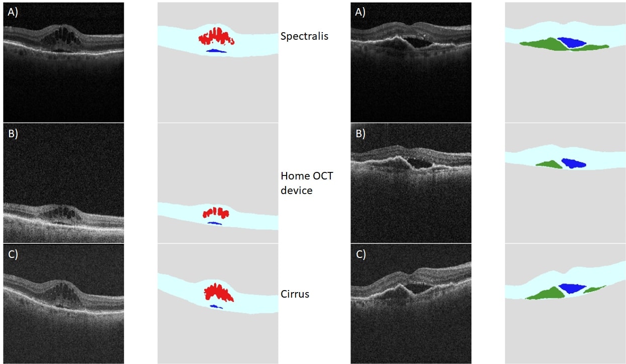
Professor Giovanni Staurenghi presented collaborative work on the “Automated quantitative assessment of neovascular age-related macular degeneration ocular coherence tomography (OCT) images acquired using a home-based OCT device” at today’s session on neovascular AMD. The work details the performance of a prototype home OCT device used to capture 136 subject eyes with neovascular AMD. Voxeleron’s analysis tools were used to segment and quantify the retinal layers as well as intra-retinal, sub-retinal and fluid beneath the retinal pigment epithelium (RPE) layer. The segmentation was run on all three devices – the prototype home device and two clinical devices (see example slide below) – and the performance was deemed akin to human graders with image quality on a par to the clinical devices. The conclusions were:
- Automated analysis of the images acquired using the prototype home OCT device achieved a high degree of accuracy for retinal layer segmentation, retinal fluid detection, and retinal fluid quantification across all retinal fluid types in comparison with manually generated measurements
- This is an important foundation for future studies of the prototype home OCT device and highlights its potential to detect the need for anti-VEGF retreatment outside the clinic
Further information can be found here:
https://www.xcdsystem.com/maculasociety/program/Na9Oa7u/index.cfm