Orion Workflow Advantages
Orion is a feature packed research tool supporting OCT analysis. Its key features are:
- Device independence – same algorithm for all devices.
- State of the art analysis tools.
- Multi-layer segmentation.
- Angiography quantification.
- Longitudinal analysis.
- Best in class editing, annotation and visualization tools.
But what is sometimes overlooked is how impactful this is with respect to workflow and time to results. If we break down each of Orion’s components, how does this impact, for example, a clinical trial?
Device Independence
Orion unifies OCT analysis by supporting all OCT devices and their formats with a common algorithm. This means that data from different devices can be analyzed and compared using Orion – allowing the reader to avoid alternating between different software applications to review endpoints. This also means that recruitment for the trial can be simplified and sped up as a single OCT device is not required. With faster recruitment, faster analysis, and more and better endpoints, clinical trials using Orion can conclude earlier – representing significant cost savings in the development of new therapeutics. Furthermore, in supporting all devices and their formats, our DICOM export functionality offers a conduit to existing IT infrastructure that simply would not otherwise exist.

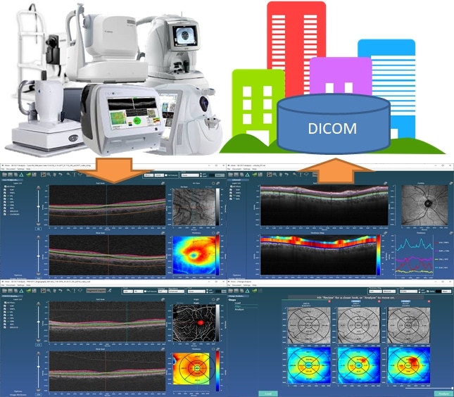
Orion is capable of reading data from all OCT devices, performing analysis and then exporting results and image data to standard DICOM (image data using lossless compression).
State of the Art Analysis Tools
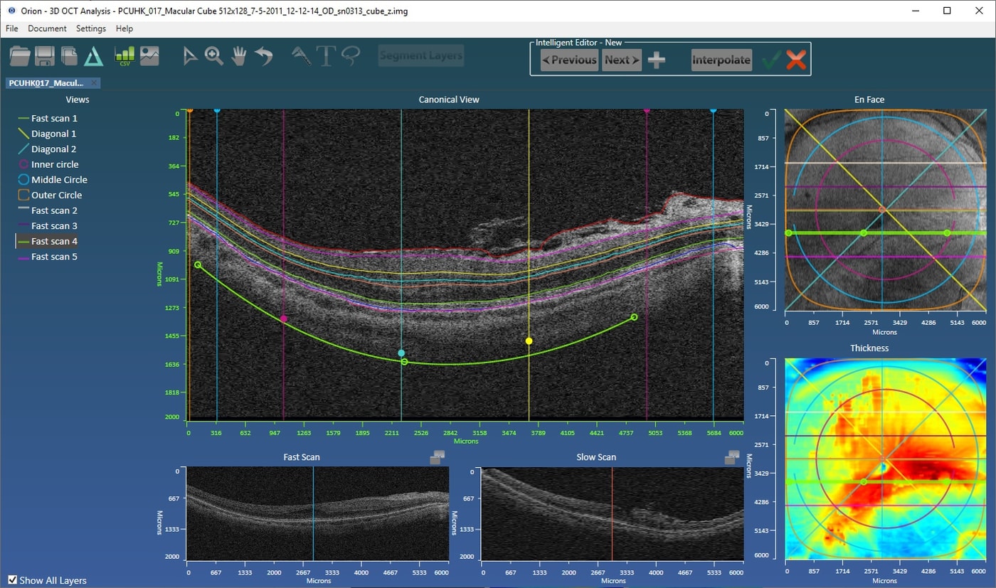
The analysis software that ships with the devices is extremely limited in terms of functionality. Being research software, Orion has continually evolved to offer best in class analysis tools validated with multiple journal publications. If you are monitoring, for example, drusen volume over time, there is no better solution than Orion. Multiple layer segmentation also supports accurate definition of the different retinal vascular plexuses, which in turn offers more accurate angiography quantification. The workflow is not only fast, it offers more insights into the OCT data. And this can all be automated using batch processing!

Editing, Annotation and Visualization
All data that is used in a trial must be quality controlled and approved by a reader who needs to be able to record each endpoint as quickly and as accurately as possible. So the automated analysis must be fast, and the review tools intuitive and simple. This includes the ability to delineate regions and add calipers to the image data. All summary results formats need to be supported (ETDRS, quadrants and ellipsoidal annuli) and, when necessary, layer editing should be simple and fast. Our patent-pending, intelligent editing wizard automates custom views through the volumetric data, allowing the user to address just what is needed and let the algorithms do the rest.

More information can be acquired watching our video tutorials, or just get in touch and request a trial!
OPHTHALMIC LINKS
Journal Publications
“Corneal Nerve Changes Observed by In Vivo Confocal Microscopy in Patients Receiving Oxaliplatin for Colorectal Cancer: The COCO Study”, Tyler, McGhee, Lawrence et al. J. Clin. Med. 2022, 11(16), 4770.
https://www.mdpi.com/2077-0383/11/16/4770
“Utilization of deep learning to quantify fluid volume of neovascular age-related macular degeneration patients based on swept-source OCT imaging: The ONTARIO study”, Simrat K. Sodhi, Austin Pereira, Jonathan D. Oakley et al. PLOS ONE 17(2): e0262111, 2022.
https://doi.org/10.1371/journal.pone.0262111
“Assessing the validity of a cross‑platform retinal image segmentation tool in normal and diseased retina”, Alex, V., Motevasseli, T., Freeman, W.R. et al. Sci Rep 11, 21784 (2021). https://doi.org/10.1038/s41598-021-01105-9.
https://www.nature.com/articles/s41598-021-01105-9
“Corneal Confocal Microscopy Demonstrates Axonal Loss in Different Courses of Multiple Sclerosis”, Petropoulos, Fitzgerald, Oakley et al. Scientific Reports volume 11, Article number: 21688 (2021).
https://www.nature.com/articles/s41598-021-01226-1
“Automated Deep Learning-Based Multi-Class Fluid Segmentation in Swept-Source Optical Coherence Tomography Images”, Jonathan D Oakley, Simrat K Sodhi, Daniel B Russakoff and Netan Choudhry. EC Ophthalmology 12.8 (2021): 24-37.
https://www.ecronicon.com/ecop/ECOP-12-00792.php
“Longitudinal Study of Retinal Structure, Vascular, and Neuronal Function in Patients With Relapsing-Remitting Multiple Sclerosis: 1-Year Follow-Up.”, Chen Q, Jiang H, Delgado S et al. Trans. Vis. Sci. Tech. 2021;10(6):6. doi: https://doi.org/10.1167/tvst.10.6.6.
https://tvst.arvojournals.org/article.aspx?articleid=2772568
“Effects of subthreshold nanosecond laser therapy in age-related macular degeneration using artificial intelligence (STAR-AI Study)”, Hanna V, Oakley J, Russakoff D, Choudhry N. PLoS ONE 16(4): e0250609. https://doi.org/10.1371/journal.pone.0250609
https://journals.plos.org/plosone/article?id=10.1371/journal.pone.0250609
“Combining In Vivo Corneal Confocal Microscopy With Deep Learning-Based Analysis Reveals Sensory Nerve Fiber Loss in Acute Simian Immunodeficiency Virus Infection. Cornea”, McCarron ME, Weinberg RL, Izzi JM, Queen SE, Tarwater PM, Misra SL, Russakoff DB, Oakley JD, Mankowski JL. Cornea: May 2021 – Volume 40 – Issue 5 – p 635-642.
“Inner retinal thickening affects microperimetry thresholds in the presence of photoreceptor thinning in patients with RPGR retinitis pigmentosa”, Jolly JK, Menghini M, Johal PA, et al. British Journal of Ophthalmology Published Online First: 30 October 2020.
https://bjo.bmj.com/content/early/2020/10/30/bjophthalmol-2020-317692
https://www.biorxiv.org/content/10.1101/2020.09.01.278259v1
“Deep Learning-Based Analysis of Macaque Corneal Sub-Basal Nerve Fibers in Confocal Microscopy Images”, Jonathan D. Oakley, Daniel B. Russakoff, Megan E. McCarron, Rachel L. Weinberg, Jessica M. Izzi, Stuti L. Misra, Charles N. McGhee, Joseph L. Mankowski. Eye and Vision (2020) 7:27 https://doi.org/10.1186/s40662-020-00192-5.
https://eandv.biomedcentral.com/articles/10.1186/s40662-020-00192-5
“Combining In Vivo Corneal Confocal Microscopy with Deep Learning-based Analysis Reveals Sensory Nerve Fiber Loss in Acute SIV Infection”, ME McCarron, RL Weinberg, JM Izzi, SE Queen, SL Misra, DB Russakoff, J Oakley, J Mankowski. bioRxiv doi: https://doi.org/10.1101/2020.04.19.048926
https://doi.org/10.1101/2020.04.19.048926
“Focal alteration of the intraretinal layers in neurodegenerative disorders”, Airen S, Shi C, Liu Z, Levin BE, Signorile JF, Wang J and Jiang H. Annals of Eye Science, Vol. 5, 2020.
http://aes.amegroups.com/article/view/5193
“A 3D Deep Learning System for Detecting Referable Glaucoma Using Full OCT Macular Cube Scans”, Daniel B. Russakoff; Suria S. Mannil; Jonathan D. Oakley; et al. Translational Vision Science & Technology February 2020, Vol.9, 12.
https://tvst.arvojournals.org/article.aspx?articleid=2761680
“Association of longitudinal changes in drusen characteristics and retinal layer volumes with subsequent subtype of choroidal neovascularisation”, Lamin A, El Nokrashy A, Chandra S, Sivaprasad S. Ophthalmic Res 2019. doi: 10.1159/000505628
https://www.karger.com/Article/Abstract/505628#
“Retinal thinning of inner sub-layers is associated with cortical atrophy in a mouse model of Alzheimer’s disease: a longitudinal multimodal in vivo study”, Chiquita, S., Campos, E.J., Castelhano, J. et al. Alz Res Therapy (2019) 11: 90.
https://doi.org/10.1186/s13195-019-0542-8
“Focal Thickness Reduction of the Ganglion Cell-Inner Plexiform Layer Best Discriminates Prior Optic Neuritis in Patients With Multiple Sclerosis”, Huiling Hu; Hong Jiang; Giovana Rosa Gameiro et al. Investigative Ophthalmology & Visual Science October 2019, Vol.60, 4257-4269.
https://iovs.arvojournals.org/article.aspx?articleid=2753187
“Deep Learning-Based Analysis of Macaque Corneal Sub-Basal Nerve Fibers in Confocal Microscopy Images”, Jonathan D Oakley, Daniel B Russakoff, Rachel L Weinberg, Megan E McCarron, Jessica M Izzi, Joseph L Mankowski. bioRxiv
https://www.biorxiv.org/content/10.1101/758433v1
“Inner Nuclear Layer Microcyst Configuration, Distribution, and Visual Prognosis in Patients With Epiretinal Membrane After Vitrectomy and Membrane Peeling”, Ming-Hung Hsieh, Yu-Bai Chou, Yi-Ming Huang, De-Kuang Hwang, Fang-Yi Tsai & Shih-Jen Chen. Nature Scientific Reports, Volume 9, Article number: 11570 (2019).
https://www.nature.com/articles/s41598-019-48097-1
“Visual Function and Disability Are Associated With Focal Thickness Reduction of the Ganglion Cell-Inner Plexiform Layer in Patients With Multiple Sclerosis”, Ce Shi; Hong Jiang; Giovana Rosa Gameiro; Huiling Hu; Jeffrey Hernandez; Silvia Delgado; Jianhua Wang. Investigative Ophthalmology & Visual Science March 2019, Vol.60, 1213-1223.
https://iovs.arvojournals.org/article.aspx?articleid=2729572
“Age-related Alterations in Retinal Tissue Perfusion and Volumetric Vessel Density”, Ying Lin, Hong Jiang, Yi Liu, Giovana Rosa Gameiro, Giovanni Gregori, Chuanhui Dong, Tatjana Rundek, and Jianhua Wang. Invest Ophthalmol Vis Sci. February 2019, Vol.60, 685-693.
https://iovs.arvojournals.org/article.aspx?articleid=2725781
“Deep Learning for Prediction of AMD Progression: A Pilot Study”, Daniel B. Russakoff, Ali Lamin, Jonathan D. Oakley, Adam M. Dubis, and Sobha Sivaprasad. Invest Ophthalmol Vis Sci. February 2019, Vol.60, 712-722.
https://iovs.arvojournals.org/article.aspx?articleid=2725851
“Changes in volume of various retinal layers over time in early and intermediate age-related macular degeneration”, Ali Lamin, Jonathan D. Oakley, Adam M. Dubis, Daniel B. Russakoff & Sobha Sivaprasad. Eye (2018)
https://www.nature.com/articles/s41433-018-0234-9
“Visualization of Focal Thinning of the Ganglion Cell–Inner Plexiform Layer in Patients with Mild Cognitive Impairment and Alzheimer’s Disease”, Y Shao, H Jiang, Y Wei et al. Journal of Alzheimer’s Disease, vol. 64, no. 4, pp. 1261-1273, 2018.
https://content.iospress.com/articles/journal-of-alzheimers-disease/jad180070
“Predictors of Retinal Atrophy in Multiple Sclerosis: A Longitudinal Study Using Spectral Domain Optical Coherence Tomography with Segmentation Analysis”, Raed Behbehani, Hussain Adnan, Abdullah Abu Al-Hassan, Ali Al-Salahat and Raed Alroughani.
Mult Scler Relat Disord. February, 2018.
http://www.msard-journal.com/article/S2211-0348(18)30055-5/fulltext
“Retinal layers thickness changes following epiretinal membrane surgery”, I Hecht, I Yeshurun, E Bartov, A Bar, Z Burgansky-Eliash & A Achiron. Eye, Nov 2017, 10.1038/eye.2017.233.
https://www.nature.com/articles/eye2017233
“Age-Related Alterations in the Retinal Microvasculature, Microcirculation, and Microstructure”, Wei Y, Jiang H, Shi Y, Qu D, Gregori G, Zheng F, Rundek T, Wang J. Invest Ophthalmol Vis Sci. 2017 Jul 1;58(9):3804-3817.
http://iovs.arvojournals.org/article.aspx?articleid=2646459
“Increased susceptibility to fundus camera-delivered light-induced retinal degeneration in mice deficient in oxidative stress response proteins”, Ding Y, Aredo B, Zhong X, Zhao CX, Ufret-Vincenty RL. Experimental Eye Research, 2017 Jun;159:58-68.
https://www.ncbi.nlm.nih.gov/pubmed/28336262#
“Simultaneous Fluorescein Angiography and SDOCT Correlate Retinal Thickness Changes to Vascular Abnormalities in an In Vivo Mouse Model of Retinopathy of Prematurity”, Olachi J. Mezu-Ndubuisi, Lauren K. Taylor, and Jamee A. Schoephoerster. Journal of Ophthalmology, Volume 2017.
https://www.hindawi.com/journals/joph/2017/9620876/
“Optical coherence tomography segmentation analysis in relapsing remitting versus progressive multiple sclerosis”, Raed Behbehani, Abdullah Abu Al-Hassan, Ali Al-Salahat, Devarajan Sriraman, J. D. Oakley, Raed Alroughani. PLoS ONE 12(2): e0172120. doi:10.1371/journal.pone.0172120.
http://journals.plos.org/plosone/article?id=10.1371/journal.pone.0172120
“The measurement repeatability using different partition methods of intraretinal tomographic thickness maps in healthy human subjects”, Tan J, Yang Y, Jiang H, Liu C, Deng Z, Lam BL, Hu L, Oakley J, Wang J. Clinical Ophthalmology, Volume 2016:10 Pages 2403—2415, Nov 2016.
https://www.dovepress.com/articles.php?article_id=30257
“Changes in macular layers in the early course of non-arteritic ischaemic optic neuropathy”, Johannes Keller, Jonathan D. Oakley, Daniel B. Russakoff, Magí Andorrà-Inglés, Pablo Villoslada, Bernardo F. Sánchez-Dalmau. Graefe’s Archive for Clinical and Experimental Ophthalmology, DOI 10.1007/s00417-015-3066-3, May 2015.
http://9nl.us/cli_exp_ophth_aion
“Loss of Corneal Sensory Nerve Fibers in SIV-infected Macaques: An Alternate Approach to Investigate HIV-induced PNS Damage,” by Jamie L. Dorsey, Lisa M. Mangus, Jonathan D. Oakley, Sarah E. Beck, Kathleen M. Kelly, Suzanne E. Queen, Kelly A. Metcalf Pate, Robert J. Adams, Carl F. Marfurt, and Joseph L. Mankowski. The American Journal of Pathology, Volume 184, Issue 6 (June 2014) published by Elsevier.
http://ajp.amjpathol.org/article/S0002-9440(14)00162-X/fulltext
“Active MS is associated with accelerated retinal ganglion cell/inner plexiform layer thinning”, John N. Ratchford, MD, Shiv Saidha, MRCPI, Elias S. Sotirchos, MD, Jiwon A. Oh, MD, FRCPC, Michaela A. Seigo, ScB, Christopher Eckstein, MD, Mary K. Durbin, PhD, Jonathan D. Oakley, PhD, Scott A. Meyer, PhD, Amy Conger, COA, Teresa C. Frohman, BS, Scott D. Newsome, DO, Laura J. Balcer, MD, MSCE, Elliot M. Frohman, MD, PhD and Peter A. Calabresi, MD. Neurology January 1, 2013 vol. 80 no. 1 47-54.
http://www.neurology.org/content/80/1/47
“Relationships Between Retinal Axonal and Neuronal Measures and Global Central Nervous System Pathology in Multiple Sclerosis”, Shiv Saidha, MBBCh, MRCPI; Elias S. Sotirchos, MD; Jiwon Oh, MD, FRCPC; Stephanie B. Syc, ScB; Michaela A. Seigo, ScB; Navid Shiee, MS; Chistopher Eckstein, MD; Mary K. Durbin, PhD; Jonathan D. Oakley, PhD; Scott A. Meyer, PhD; Teresa C. Frohman, BS; Scott Newsome, DO; John N. Ratchford, MD; Laura J. Balcer, MD, MSCE; Dzung L. Pham, PhD; Ciprian M. Crainiceanu, PhD; Elliot M. Frohman, MD, PhD; Daniel S. Reich, MD, PhD; Peter A. Calabresi, MD. Arch Neurol. 2012;():1-10. doi:10.1001/archneurol.2013.573.
http://www.ncbi.nlm.nih.gov/pubmed/23318513
“In vivo assessment of retinal neuronal layers in multiple sclerosis with manual and automated optical coherence tomography segmentation techniques”, Michaela A. Seigo, Elias S. Sotirchos, Scott Newsome, Aleksandra Babiarz, Christopher Eckstein, E’Tona Ford, Jonathan D. Oakley, Stephanie B. Syc, Teresa C. Frohman and John N. Ratchford, et al., Journal of Neurology 2012, DOI: 10.1007/s00415-012-6466-x.
http://www.springerlink.com/content/f03715555760813l/
“Optical coherence tomography segmentation reveals ganglion cell layer pathology after optic neuritis”, Stephanie B. Syc, Shiv Saidha, Scott D. Newsome, John N. Ratchford, Michael Levy, E’Tona Ford, Ciprian M. Crainiceanu, Mary K. Durbin, Jonathan D. Oakley, Scott A. Meyer, Elliot M. Frohman and Peter A. Calabresi. Brain (2011) doi: 10.1093/brain/awr264, First published online: October 17, 2011.
http://brain.oxfordjournals.org/content/early/2011/10/17/brain.awr264.abstract
“Macular Ganglion Cell-Inner Plexiform Layer: Automated Detection and Thickness Reproducibility with Spectral-Domain Optical Coherence Tomography in Glaucoma”, Jean-Claude Mwanza, Jonathan D. Oakley, Donald L. Budenz, Robert T. Chang, O’Rese J. Knight and William J. Feuer, Invest. Ophthalmol. Vis. Sci. September 14, 2011 iovs.11-7962.
http://www.iovs.org/content/52/11/8323.abstract
“Visual dysfunction in multiple sclerosis correlates better with optical coherence tomography derived estimates of macular ganglion cell layer thickness than peripapillary retinal nerve fiber layer thickness”, Shiv Saidha, Stephanie B. Syc, Mary K Durbin, Christopher Eckstein, Jonathan D. Oakley et al. Multiple Sclerosis Journal, August 24, 2011.
http://msj.sagepub.com/content/early/2011/08/19/1352458511418630.abstract?papetoc
“Comparison of Automated Analysis of Cirrus HD OCT Spectral-Domain Optical Coherence Tomography with Stereo Photographs of the Optic Disc”, Ophthalmology Volume 118, Issue 7, Pages 1348-1357, July 2011.
http://www.ophsource.org/periodicals/ophtha/article/PIIS0161642010012820/abstract?rss=yes
“Ability of Cirrus HD-OCT Optic Nerve Head Parameters to Discriminate Normal from Glaucomatous Eyes”, Jean-Claude Mwanza, MD, PhD, Jonathan D. Oakley, PhD, Donald L. Budenz, MD, MPH, Douglas R. Anderson, Ophthalmology Volume 118, Issue 2 , Pages 241-248.e1, February 2011.
http://www.ophsource.org/periodicals/ophtha/article/S0161-6420(10)00708-6/abstract
“Primary retinal pathology in multiple sclerosis as detected by optical coherence tomography”, Shiv Saidha, Stephanie B. Syc, Mohamed A. Ibrahim, Christopher Eckstein, Christina V. Warner, Sheena K. Farrell, Jonathan D. Oakley, Mary K. Durbin, Scott Meyer, Laura J. Balcer, Elliot M. Frohman, Jason M. Rosenzweig, Scott D. Newsome, John N. Ratchford, Quan D. Nguyen and Peter A. Calabresi, Brain, 2011 – Oxford Univ Press.
Association for Research in Vision and Ophthalmology Presentations
“Machine Learning Quantification of Fluid Volume in Eyes with Retinal Vein Occlusion Undergoing Treatment with Aflibercept: The REVOLT study”, Mohammad Khan, Simrat Sodhi, John Golding, Austin Pereira, Anuradha Dhawan, Jonathan Oakley, Daniel Russakoff, Netan Choudhry. ARVO Meeting Abstracts, 2022.
https://arvo2022.arvo.org/meetings/4zcXrSuJySCtKqzZF
“Automated segmentation of retinal nerve fiber layer excluding retinal blood vessels: integrating OCT and OCT Angiography”, Matteo Airaldi, Jonathan Oakley, Sara Bochicchio, Angelica Dipinto, Simona Prandoni, Giovanni Staurenghi, Giacinto Triolo. ARVO Meeting Abstracts, 2022.
https://arvo2022.arvo.org/meetings/onaxYQ3cEwsYbeeoh
“Proof of concept analysis of a deep learning model to conduct automated segmentation of optical coherence tomography images for macular hole volume”, Austin Pereira, Jonathan Oakley, Simrat Sodhi, Netan Choudhry. ARVO Meeting Abstracts, 2022.
https://arvo2022.arvo.org/meetings/v47NY3iuoNGmCZK94
“Utilisation of Machine Learning to quantify fluid volume of wet age-related macular degeneration (wARMD) patients based on swept-source optical coherence tomography (SS-OCT) imaging: The ONTARIO Study”, Simrat K. Sodhi, Jonathan D. Oakley, Daniel B. Russakoff and Netan Choudhry.
https://arvo2021.arvo.org/meetings/virtual/CR9xEBTHn4nujgdRf
“Assessing the validity of a cross-platform retinal image segmentation tool in normal and diseased retina”, Varsha Alex, Tahmineh Motevasseli, Jefy A. Jayamon, Sumit R. Singh, Dirk U. Bartsch, Lingyun Cheng, Shyamanga Borooah and William R. Freeman.
https://arvo2021.arvo.org/meetings/virtual/WwSRSR4NX2WmmtdXF
“Automated choroid segmentation of SD-OCT volumes using deep learning.” Oakley, Jonathan D. and Russakoff, Daniel B. ARVO Imaging in the Eye Meeting Abstracts, 2019.
https://iovs.arvojournals.org/article.aspx?articleid=2748189
“Diagnostic Assessment of RNFL Segmentation using a Hybrid Deep Learning Approach.” Oakley, Jonathan D., Mannil, Suria S.; Russakoff, Daniel; Chang, Robert. ARVO Meeting Abstracts, 2019.
https://iovs.arvojournals.org/article.aspx?articleid=2747025&resultClick=1
“A 3D Deep Learning System for Detecting Referrable Glaucoma Using Full OCT Macular Cube Scans.” Russakoff, Daniel; Mannil, Suria S.; Oakley, Jonathan D.; Chang, Robert. ARVO Meeting Abstracts, 2019.
https://iovs.arvojournals.org/article.aspx?articleid=2747085&resultClick=1
“Novel deep learning based algorithm for Macula and Optic Nerve Head segmentation versus Cirrus Optical Coherence Tomography in identifying glaucoma.” Sudhakaran Mannil, Suria; Oakley, Jonathan D.; Russakoff, Daniel B.; Chang, Robert. ARVO Meeting Abstracts, 2019.
https://iovs.arvojournals.org/article.aspx?articleid=2746565&resultClick=1
“Deep learning for prediction of AMD progression”, Daniel Russakoff, Ali Lamin, Jonathan Oakley, Adam Dubis, Susan Lightman, Sobha Sivaprasad. ARVO Meeting Abstracts, 2018.
https://iovs.arvojournals.org/article.aspx?articleid=2694276&resultClick=1
“Automated Analysis of In Vivo Confocal Microscopy Corneal Images Using Deep Learning”, Jonathan Oakley, Daniel Russakoff, Rachel Weinberg, Megan McCarron, Samuel Brill, Stuti Misra, Charles McGhee, Joseph Mankowski. ARVO Meeting Abstracts, 2018.
https://iovs.arvojournals.org/article.aspx?articleid=2694279&resultClick=1
“Relevance and Validation of Optical Coherence Tomography based on Volumetric Measures in Age-Related Macular Degeneration”, Ali Lamin, Jonathan Oakley, Adam Dubis, Susan Lightman, Sobha Sivaprasad. ARVO Meeting Abstracts, 2018.
https://iovs.arvojournals.org/article.aspx?articleid=2693768&resultClick=1
“Retinal microcirculation, microvasculature and microstructure in patients with multiple sclerosis: 1 year follow-up”, Hong Jiang, Ce Shi, Zhengyu Duan, Silvia Delgado, Giovanni Gregori, Jeffrey Hernandez, Jianhua Wang. ARVO Meeting Abstracts, 2018.
https://iovs.arvojournals.org/article.aspx?articleid=2689362
“Interaction between retinal microcirculation and microstructure in patients with Alzheimer’s disease”, Ying Lin, Hong Jiang, Yi Liu, Yuqing Deng, Zhengyu Duan, Tatjana Rundek, Xiaoyan Sun, Bernard Baumel, Jianhua Wang. ARVO Meeting Abstracts, 2018.
https://iovs.arvojournals.org/article.aspx?articleid=2693345
“Age-related focal thinning of the ganglion cell–inner plexiform layer”, Jianhua Wang, Yuqing Deng, Ce Shi, Tatjana Rundek, Bernard Baumel, Hong Jiang. ARVO Meeting Abstracts, 2018.
https://iovs.arvojournals.org/article.aspx?articleid=2693342
“Deep convolutional neural networks for automated OCT pathology recognition”, Russakoff, Daniel B., Oakley, Jonathan D., Chang, Robert. ARVO Meeting Abstracts, 2017.
https://iovs.arvojournals.org/article.aspx?articleid=2637970&resultClick=1
“Automated analysis of in vivo confocal microscopy images of corneal nerves”, Stuti L Misra, Jonathan D Oakley, Charles N McGhee, 1 Ellen F Wang, Dipika V Patel, Patrick M Tarwater, Joseph L Mankowski. ARVO Meeting Abstracts, 2017.
https://iovs.arvojournals.org/article.aspx?articleid=2637559&resultClick=1
“Comparison of Automated Retinal Segmentation using Different Optical Coherence Tomography Devices” – Jonathan D. Oakley, Magí Andorrà Inglés, Elena H. Martínez-Lapiscina, Daniel Russakoff, and Pablo Villoslada. ARVO Meeting Abstracts, 2016.
http://www.arvo.org/webs/am2016/sectionpdf/MOI/Session_514.pdf
“Correlation of Intraretinal Thickness and Microvasculature in Healthy Subjects” – Hong Jiang, Min Li, Jin Zhou, Ye Yang, Wan Chen, Liang Hu and Jianhua Wang. ARVO Meeting Abstracts, 2016.
http://www.arvo.org/webs/am2016/sectionpdf/PH/Session_420.pdf
“Relationship between Axial Length and Macula Retinal layer thickness in Normal Human Subjects”, Kwame Antwi-Boasiako, Jonathan D. Oakley, Daniel Russakoff, Sherine John, Nimesh B. Patel. ARVO Meeting Abstracts, 2015.
http://www.arvo.org/webs/am2015/abstract/503.pdf
“Assessing Manual versus Automated Segmentation of the Macula using Optical Coherence Tomography”, Jonathan D. Oakley, Iñigo Gabilondo, Christopher Songster, Daniel Russakoff, Ari Green, Pablo Villoslada. ARVO Meeting Abstracts, 2014.
http://iovs.arvojournals.org/article.aspx?articleid=2270338
“True Outer Nuclear Layer Volumes Using Directional Optical Coherence Tomography”, Brandon J. Lujan, Daniel Russakoff, Jonathan D. Oakley, Mona K. Garvin, Austin Roorda. ARVO Meeting Abstracts, 2014.
http://iovs.arvojournals.org/article.aspx?articleid=2270353
“Analyzing Shape Parameterization of SD-OCT Optic Nerve Head Images in High Myopes as a Predictor of Visual Field Defects”, Sonny Sabhlok, Daniel Russakoff, Tessa Johung, Jonathan Oakley, Felix Li, Kuldev Singh, Robert Chang, ARVO Meeting Abstracts, 2013.
http://www.arvo.org/webs/am2013/abstract/sessions/231.pdf
“The Prevalence of Cirrus SD-OCT Ganglion Cell Segmentation Errors in High Myopes”, Tessa Johung, Jonathan Oakley, Daniel Russakoff, Sonny Sabhlok, Felix Li, Robert Chang, ARVO Meeting Abstracts, 2013.
http://www.arvo.org/webs/am2013/abstract/sessions/443.pdf
“Diagnostic performance of Cirrus HD-OCT Ganglion Cell-Internal Plexiform Layer Measurements to Discriminate Between Normal and Glaucomatous Eyes”, FE Sayyad, JC Mwanza, JD Oakley, DL Buden. ARVO Meeting Abstracts, 2011.
http://abstracts.iovs.org/cgi/content/short/52/6/185
“Ability of CirrusTM HD-OCT Optic Disc Measurements to Discriminate Between Normal and Glaucomatous Eyes”, J.-C. Mwanza, J.D. Oakley, D.R. Anderson, D.L. Budenz. ARVO Meeting Abstracts, 2010.
“Effect of Ethnicity, Age, and Axial Length on Optic Nerve Head Parameters Measured by CirrusTM HD- OCT”, O.J. Knight, J.D. Oakley, M. Durbin, T. Callan, D.L. Budenz, Cirrus Normative Database Study Group. ARVO Meeting Abstracts, 2010.
“Reproducibility of Optic Nerve Head Parameters Measured With Cirrus HD-OCT in Glaucomatous Eyes”, R.T. Chang, J.-C. Mwanza, M.G. Gendy, J.D. Oakley, W.J. Feuer, D.L. Budenz. ARVO Meeting Abstracts, 2010.
“Comparison of Evaluation of the Optic Nerve by Use of Stereo Color Photos and Spectral Domain Optical Coherence Tomography”, A. Sharma, J.D. Oakley, J Schiffman, D.L. Budenz, D.R. Anderson. ARVO Meeting Abstracts, 2010.
“Comparing Optic Nerve Head Parameters from the Cirrus HD-OCT and Stratus OCT”, TA Abunto, M Durbin, J Oakley. ARVO Meeting Abstracts, 2010.
“Effect of Image Registration on Repeatability of CirrusTM HD-OCT Thickness Measurements”, H. Narasimha-Iyer, M. Durbin, J. Oakley, M. Everett, T. Callan, M. Horne, T. Abunto. ARVO Meeting Abstracts, 2009.
“Improved Repeatability Using Automated Post-Acquisition Disc Centering on Images From CirrusTM HD-OCT”, M. Durbin, S. Meyer, S. Dastmalchi, and J. Oakley, ARVO Meeting Abstracts April 11, 2008 49:4622
http://abstracts.iovs.org//cgi/content/abstract/49/5/4622?sid=f8ccadcd-ec29-4f7c-8a29-a4967f8af46f
“Registration of Cirrus HD-OCT Images With Fundus Photographs, Fluorescein Angiographs and Fundus Autofluorescence Images”, H. Narasimha-Iyer, B. Lujan, J. Oakley, S. Meyer, and S. S. Dastmalchi, ARVO Meeting Abstracts April 11, 2008 49:1831
White Papers
“Regulatory Aspects of Releasing OCT Normative Databases and Algorithms”, Jonathan D Oakley.
https://voxeleron.com/resources/
“Algorithms for Clinical Optical Coherence Tomography”, Jonathan D Oakley.
https://voxeleron.com/resources/
“Optic Nerve Head Normative Values”, Jonathan D Oakley, Mark K Durbin and Tom Callan.
Other Conference Presentations
“Machine Learning Quantification of Fluid Volume in Eyes with Neovascular Age-Related Macular Degeneration and Retinal Vein Occlusion: The ONTARIO and REVOLT studies”, Sodhi, Khan, Golding et al. Euretina 2022.
“A comparison between two automated algorithms to segment the inner and outer retinal thicknesses in eyes with neovascular age-related macular degeneration.”, Tibaldi, Caselgrandi, Oakley et al. Euretina 2022.
“Retinal Neurodegeneration and retinal vessel changes assessed by Optical Coherence Tomography Angiography in type 1 Diabetes Mellitus. A large scale OCTA study”, Castell, Rosines-Fonoll, Martin-Pinardel et al. Euretina 2022.
“Quantitative Assessment of Automated Ocular Coherence Tomography (OCT) Image Analysis Using a Home-Based OCT Device for Self-Monitoring Neovascular Age-related Macular Degeneration”, Giovanni Staurenghi, Steven Verdooner, Jonathan Oakley et al. 45th Annual Macula Society Meeting in Berlin, Germany, June 2022.
https://www.xcdsystem.com/maculasociety/program/Na9Oa7u/index.cfm
“Automated segmentation of retinal nerve fiber layer excluding peripapillary blood vessels: integrating OCT and OCT Angiography”, Valentina Folegani, Matteo Airaldi, Jonathan D Oakley, Sara Bochicchio, Angelica Dipinto, Simona Prandoni, Giovanni Staurenghi, Giacinto Triolo. European Glaucoma Society 15th Congress, Athens June 2022.
“Deep-Learning based Automated Quantification of Critical OCT Features in Neovascular Age-related Macular Degeneration”, Borrelli, Oakley, Grosso et al. Euretina 2021.
“Proof of Concept Validation of an Artificial Intelligence Model to Conduct Automated Segmentation of Swept-Source Optical Coherence Tomography Images for Macular Hole Volume” Austin Pereira, Jonathan Oakley, Netan Choudhry. 54th Annual Scientific Meeting of the Retina Society, Chicago, 2021.
https://www.retinasociety.org/content/documents/posters_round2.pdf
“Automated diabetic macular edema screening in primary care with spectral domain optical coherence tomography”, M. Carrión Donderis, A. Sala Puigdollers, M. Figueras Roca, B. Sanchez Dalmau, A. Adán, J. Zarranz Ventura. Euretina 2019.
http://www.euretina.org/downloads/EURETINA_Paris19%20Final%20Programme.pdf
“Mapping focal loss of the ganglion cell–inner plexiform layer in patients multiple sclerosis”, Jianhua Wang, Hong Jiang, Silvia Delgado, Ce Shi, Yi Shao, Yingying Shi. ECTRIMS Online Library. Wang J. Oct 26, 2017; 200057
“Signal quality dependency of intra-retinal segmentation algorithms in macular optical coherence tomography” T Oberwahrenbrock, R Jost, H Zimmermann, I Beckers, F Paul, A.U Brandt. ECTRIMS Online Library. Oberwahrenbrock T. Sep 15, 2016; 146399.
“A Window to the Peripheral Nervous System: Corneal Sensory Nerve Fiber Loss in SIV-infected Macaques”, L Mangus, J Dorsey, J Oakley, J Mankowski. JOURNAL OF NEUROVIROLOGY 19, S53-S53
“Vitreo-Retinal Interface Segmentation from Spectral-Domain OCT Using Change Detection and Belief Propagation”, Wenmiao Lu, Jonathan Oakley, Daniel Russakoff and Robert Chang, ISBI 2013. Proceedings, in press, (2013)
“Clinical and Radiological Disease Activity Is Associated with Accelerated Rates of Retinal Ganglion Cell Layer Degeneration in Multiple Sclerosis”, Shiv Saidha, John Ratchford, Elias Sotirchos, Christopher Eckstein, Jonathan Oakley, Mary Durbin, Scott Meyer, Stephanie Syc, Michaela Seigo, Scott Newsome, Laura Balcer, Elliot Frohman, Peter Calabresi, American Academy of Neurology, 2012.
“Cortical grey-matter atrophy correlates with retinal neuronal but not retinal axonal loss in multiple sclerosis”, Stephanie B. Syc, Shiv Saidha, John N. Ratchford, Navid Shiee, Michaela Seigo, Aleksandra Stankiewicz, E’Tona Ford, Mary K. Durbin , Jonathan D. Oakley, Scott A. Meyer, Elliot M. Frohman, Daniel S. Reich, Peter A. Calabresi, ECTRIMS, 2011, Amsterdam, the Netherlands.
“Retinal neuronal layer thinning and visual dysfunction in the absence of optic neuritis: evidence for subclinical disease activity in NMO”, S. Syc, S. Saidha, S. Newsome, J. Ratchford, M. Levy, J. Oakley, M. Durbin, S. Meyer, M. Seigo, A. Stankiewicz, E. Ford, E. Frohman, P. Calabresi, ECTRIMS, 2011, Amsterdam, the Netherlands.
“Primary retinal neuronal mechanisms of pathology in multiple sclerosis: challenging a paradigm”, S. Saidha, S. Syc, C. Eckstein, M. Seigo, A. Stankiewicz, E. Ford, M. Durbin, J. Oakley, S. Meyer, S. Newsome, J. Ratchford, E. Frohman, P. Calabresi, ECTRIMS, 2011, Amsterdam, the Netherlands.
“Longitudinal retinal neuronal thinning occurs in MS and is associated with visual loss and disability progression”, S. Saidha, S. Syc , M. Durbin, C. Eckstein, A. Stankiewicz, M. Seigo, E. Ford, J. Oakley, S. Meyer, S. Newsome, E. Frohman, J. Ratchford, Peter A. Calabresi, ECTRIMS, 2011, Amsterdam, the Netherlands.
http://www.congrex.ch/ectrims2011
“A Novel Method to Detect Local Ganglion Cell Layer Loss in Glaucoma by Using Spectral-Domain Optical Coherence Tomography”, K. Takayama, M. Hangai, S. Mori, M. Nukada, N. Nakano, S. Morooka, T. Akagi, A. Nonaka, H. Ikeda, J. Oakley, M. Durbin, N. Yoshimura, P264, Clinical Examination Methods, World Glaucoma Congress, Paris, 2011.
http://www.oic.it/wgc2011/pdf/abstract/P130-P324.pdf
“Primary retinal pathology in multiple sclerosis as detected by optical coherence tomography”, S. Saidha, C. Eckstein, S. Syc, C. Warner, S. Farrell, N. Shiee, P. Bazin, D. Reich, J. Oakley, M. Durbin, S. Meyer, L. Balcer, E. Frohman, J. Rosenzweig, J. Ratchford, P. Calabresi, ECTRIMS 2010, Gothenburg, Sweden.