Introducing 2d Segmentation
Introducing ZoneDetectTM
We are pleased to announce the release of ZoneDetectTM, OrionTM’s new 2d segmentation tool. ZoneDetect provides a fast, intuitive method for precise segmentations of 2D structures. Once approved by the reader, the measure is ready to be recorded and stored via iNebulaTM, where it will be secure and searchable via the world’s only ophthalmic graph database.
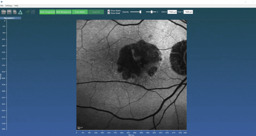
Making 2d measurements is important for endpoints derived from modalities such as fundus autofluorescence (FAF), fluorescein angiography (FA) and also color fundus photography (CFP). FAF’s signal depends on the concentration of lipofuscin in the retina, which, depending on the pathology, can result in hyper-fluorescence – for example, central serous chorioretinopathy, optic disc drusen, macular telangiectasia – or hypo-autofluorescence – for example, RPE atrophy, media opacities, intra- or sub-retinal hemorrhage. An example is given below where the RPE atrophy is a result of advanced, dry AMD, also known as geographic atrophy. Measuring the area of such atrophy is a critical endpoint in the developing new therapeutics that might slow or hopefully stop its progression, thus preserving vision. As the example below shows, recording these areas using this new tool is fast and effective. When tied to optical coherence tomography (OCT) images – using, for example, our fundus import tool – allows viewing of the fundus image with respect to the OCT data so one can see the direct correspondence across the modalities, seeing structural depth information at an precisely known position in an imported 2d image. Segmentation of regions such as the ellipsoid zone (EZ) are then supported by different modalities, facilitating exacting measurements that can be used for clinical research or as trial endpoints.

